Sub Chronic Toxicity Study of Aqueous Leaves Extract of Maytenus Gracilipes on Some Biochemical Parameters and Histopathology of Liver and Kidneys in Mice
Introduction: Traditional medicine is an ancient medical practice that is still widely used in prevention and treatment of various health problems worldwide, including Ethiopia. M.gracilipes is one of medicinal plant in Ethiopia used for treatment of various ailments still is very common. This study evaluated the sub-chronic toxic effects of M. gracilipes aqueous leaves extract on biochemical parameters and histopathology of liver and kidneys. Methods: For sub-chronic toxicity study a total of 30 mice were used, three groups (I–III) of mice (10 animals each) were used. Group I served as control and received a vehicle while groups II and III daily administered with 700 and 2100 mg/kg extract respectively orally by using oral gavage for 90 days. At the end of the experiment, the mice were sacrificed by using diethyl ether; blood was collected for assessing biochemical parameters and histopathological evaluations on liver and kidneys were performed. Results: Sub chronic treatment of extract for 90 days, at 700 and 2100 mg/kg body weight did not induce any sign of illness and /or death and had no adverse effect on biochemical parameters and blood parameters. Liver and kidney sections also revealed normal architecture, except some pyknotic nuclei and focal mononuclear leukocytic infiltrations observed in some of the liver and kidney tissues at higher dose (2100mg/kg). Conclusion: The results of this sub chronic toxicity study showed that M. gracilipes aqueous leaves extract is safe at daily doses of 700 mg/kg body weight, even when taken for longer period. At higher doses, however, the extract may induce mild hepatorenal toxicity.
This is an Open Access article, distributed under the terms of the Creative Commons Attribution 4.0 International License (http://creativecommons.org/licenses/by/4.0/), which permits unrestricted use, distribution and reproduction in any medium or format, provided the original work is properly cited.
Herbal medicine is also known as phytochemical or botanical medicine. According to World Health Organization (WHO), herbal medicine includes ‘herbs, herbal materials, herbal preparations and finished herbal products that contain active ingredient parts of plants or other plant materials or combinations there
[1]
Robinson M, Zhang X. THE WORLD MEDICINES SITUATION 2011 TRADITIONAL MEDICINES: GLOBAL SITUATION, ISSUES AND CHALLENGES. In 2011.
[1]
. Herbal medicine is generally considered as an integral part of dietary supplement. There is a growing interest in herbal medicine due to its long history of application and general belief that herbs are natural and intrinsically safe.
Traditional medicine is an ancient medical practice that is still widely used in prevention and treatment of various health problems worldwide, including Ethiopia
[2]
Addis G, Abebe DT, Genebo T, Urga K. Perceptions and practices of modern and traditional health practitioners about traditional medicine in Shirka district, Arsi zone, Ethiopia. In 2002.
[3]
Berhanu A. Book Review: Ethiopian Traditional Medicine: Common Medicinal Plants in Perspective. J Black Stud. 2002 May 1; 32(5): 610–2.
[4]
De Smet PAGM. Traditional pharmacology and medicine in Africa: Ethnopharmacological themes in sub-Saharan art objects and utensils. J Ethnopharmacol. 1998 Nov 1; 63(1): 1–175.
[5]
Gedif T, Hahn H-J. The use of medicinal plants in self-care in rural central Ethiopia. J Ethnopharmacol. 2003 Aug 1; 87(2): 155–61.
[2-5]
. There is a growing recognition that knowledge of traditional medicine is important not only for its potential as therapeutic drugs but also for its socioeconomic and cultural components
[6]
Alves RR, Rosa IM. Biodiversity, traditional medicine and public health: where do they meet? J Ethnobiol Ethnomedicine. 2007 Mar 21; 3(1): 14.
[7]
Azaizeh H, Saad B, Khalil K, Said O. The State of the Art of Traditional Arab Herbal Medicine in the Eastern Region of the Mediterranean: A Review. Evid Based Complement Alternat Med. 2006 Jun; 3(2): 229–35.
[8]
Gedif T, Hahn H-J. Herbalists in Addis Ababa and Butajira, Central Ethiopia: Mode of service delivery and traditional pharmaceutical practice. Ethiop J Health Dev. 2002 Jan 1; 16(2): 183-189–189.
[6-8]
. Before the development of synthetic or semi-synthetic medicines, folklore use of herbals was very common in rural areas, the use of herbal preparations for the treatment of various ailments is still very common
[9]
Chester AH, Yacoub MH. The role of endothelin-1 in pulmonary arterial hypertension. Glob Cardiol Sci Pract. 2014 Jun 18; 2014(2): 62–78.
[9]
. Many of the modern pharmaceuticals are derived from medicinal herbs. In theeighteenth century, when the medicinal therapy era was being introduced, the herbal treatment was the most preferred and available therapy. Many compounds from herbal origin have achieved widespread use as medicinal agents e.g. Taxols from Taxusbaccata (English yew) asanticancer agents, Silibinin from Silybummarianum (Milk thistle) as liver tonic
[10]
Kumaran RS, Jung H, Kim HJ. In vitro screening of taxol, an anticancer drug produced by the fungus, Colletotrichum capsici. Eng Life Sci. 2011; 11(3): 264–71.
[11]
Schümann J, Prockl J, Kiemer AK, Vollmar AM, Bang R, Tiegs G. Silibinin protects mice from T cell-dependent liver injury. J Hepatol. 2003 Sep 1; 39(3): 333–40.
[10, 11]
. The herbal therapyencompasses Ayurvedic, Naturopathic, Biochemical, Unani, Chinese, African and Native American medicine
[12]
Angerhofer CK. Herbal Medicines, a Guide for Healthcare Professionals, Second Edition By Joanne Barnes (University of London), Linda A. Anderson (Medicines Control Agency, London), and J. David Phillipson (University of London). Pharmaceutical Press, London, UK. 2002. xiv + 530 pp. 18.5 × 24.5 cm. $59.95. ISBN 0-85369-474-5. J Nat Prod. 2002 Dec 1; 65(12): 1964–1964.
[12]
. Herbal medicines have attained the widespreadacceptability as natural therapeutic agents for various diseases like diabetes, arthritis, renal and liver diseases, obesity and cardiovascular disorders
[13]
Mahomoodally MF. Traditional Medicines in Africa: An Appraisal of Ten Potent African Medicinal Plants [Internet]. Evidence-Based Complementary and Alternative Medicine. 2013 [cited 2019 Dec 27]. Available from:
The ethnomedical value of Maytenus gracilipes is numerous such as antioxidant, anti- inflammatory, anti- ulcer, anti- cancer and many stomach complications, antitumor, antimicrobial
[14]
Adebayo JO, Yakubu MT, Egwim EC, Owoyele VB, Enaibe BU. Effect of ethanolic extract of Khaya senegalensis on some biochemical parameters of rat kidney. J Ethnopharmacol. 2003 Sep 1; 88(1): 69–72.
[14]
.
2. Methods
2.1. Plant Collection
The leaves of Maytenus gracilipes were collected from Ankober woreda, near Atsie Minilik loge, about 42 km away from Debre Birhan, Northern Shawa, Amhara Region, 130 km North of Addis Ababa during the month of May 2016. Specimens of the plant were identified by a taxonomist and samples were deposited at the National Herbarium in the College of Natural and Computational Sciences, Addis Ababa University (AAU) for future reference with a Voucher specimen number (119/AMA/PHARM).The collected plant material was processed and extracted at phytochemical laboratory of Ethiopian Public Health Institution (EPHI).The leaves of plant were dried under shade area at room temperature and crushed, and dry powder was prepared for the study.
2.1.1. Preparation of Aqueous Leaf Extracts of Maytenus Gracilipes
The powdered leaves were macerated with distil water for 72 hrs with intermittent agitation by orbital shakerDS-500. Then, the supernatant part of agitated materials was separated from the undissolved portion of the plant material. The supernatant portion was filtered with 0.1 mm2 mesh gauze and then with Whatman grade 1 filter paper with pore size of 11𝜇m. The filtrate was then concentrated by evaporating the solvent using a rotary evaporator (BUCHIRota-vapor type R-205, Switzerland) under reduced pressure at a temperature of 40–45°C. Then the residue was dried by steambath at 40°C for period of one week to make it dry
[15]
Geyid A, Abebe D, Debella A, Makonnen Z, Aberra F, Teka F, et al. Screening of some medicinal plants of Ethiopia for their anti-microbial properties and chemical profiles. J Ethnopharmacol. 2005 Jan 1.
[15]
.
2.1.2. Preparation and Grouping of Experimental Animals
The healthy and non-pregnant young Swiss albino mice aged 8–10 weeks and weighting 25-30 gm of both sexes were obtained from EHPI, Addis Ababa. The animals were acclimated to laboratory conditions for 5 days. They were housed in standard cages and kept under standard condition at a temperature of (21+2°C) with 12hrslight/12hrs dark cycle and were provided with free access to standard diet and tap water ad libitumaccording to OECD guideline
[16]
Test No. 425: Acute Oral Toxicity: Up-and-Down Procedure - en - OECD [Internet]. [cited 2019 Jul 14]. Available from:
Animals were randomly assigned to a control and two treatment groups. Each animal was assigned a unique identification number. A total of 30 mice of both sex containing 10 mice per group (five female and five male) were used for sub chronic toxicity study.
2.2. Sub Chronic Toxicity Study
The sub chronic toxicity study was conducted for 90 days to evaluate the toxic of the extract on some biochemical parameters and histopathology of liver and kidneys by using some modification of the method used by a study
[17]
Saleem U, Amin S, Ahmad B, Azeem H, Anwar F, Mary S. Acute oral toxicity evaluation of aqueous ethanolic extract of Saccharum munja Roxb. roots in albino mice as per OECD 425 TG. Toxicol Rep. 2017 Oct 31; 4: 580–5.
[17]
.
For this study healthy adult mice of both sexes were used. Thirty mice were randomly distributed into three groups (I, II, and III) each consisting of teen mice (five female and five male) per group. Groups II and III were orally administered with aqueous extract of leaves at doses of 700 and 2100 mg/kg body weight per day respectively, for 90 days using oral gavage. Group I served as control group and received distill water. Clinical observation was carried out for 90 days and their weight was measured weekly for thirteen weeks. On the 90thday the final weight of the mice was measured and then they were anesthetized under diethyl ether and blood samples were collected from each animal by cardiac puncture.
2.3. Blood Collection for Biochemical Analyses
Blood samples in the test tubes without anticoagulant could clot and sera were obtained by centrifuging the blood using an electrical centrifuge (HUMAX-K, HUMAN-Germany)from which blood chemistry (Urea, Creatinine, Total Bilirubin, ALT, and AST) was studied to test renal and hepatic functions. Values in the sera were analyzed using Automated Clinical Chemistry Analyzer (AUTO LAB 18, clinical chemistry analyzer, Italy)
2.4. Target Organ Collection
After collection of blood samples, the mice were sacrificed by cervical dislocation and parts of the liver and the kidneys were dissected out; and gross pathological observation was performed on liver and kidneys to check for any gross lesions.
2.5. Histopathological Studies
The liver and kidney sections taken randomly for tissue processing were fixed in 10% neutral buffered formalin (NBF) overnight at room temperature. After fixation, the tissue sections were washed with water to remove excess fixatives for about six hours and dehydrated with increased concentration of alcohol of 70%for two hours, 90% for two hours, absolute alcohol-I, II for one and half-hours, and III overnight. The dehydrated tissues were cleared in two changes of xylene-for one and half hours and two and half hours. The tissues were then infiltrated with three changes of paraffin wax-for one and half hours, two and half-hours, and overnight. Finally, the tissues were embedded in paraffin wax in square metal plates forming tissue blocks, and then each tissue block was labeled and stored at room temperature till sectioned. The tissue blocks were sectioned in ribbons at a thickness of 5 𝜇m with Leica microtome (Leica RM 2125RT NusslochGmbH, Germany). The ribbons of the section were collected at every 5th sections and put onto the surface of a warm water bath of temperature of 40°C. The floating ribbons over the surface of warm water were mounted onto pre cleaned slides spread with egg albumin. The slides containing paraffin wax were arranged within the slide holder and placed in an oven with temperature of 40°C for about 20 minutes so as to fix the tissue to the slides and allowed to cool at room temperature for 30 minutes and stained regressively with routine Harrisheamatoxylin for 6 minutes and then eosin for 17-20 second (H and E). For routine H and E staining, two series of coupling jars were prepared: one for paraffin removal and hydration and the other for dehydration and clearing. So, sections were placed in xylene I for 5 minutes and xylene II for 2 minutes again to remove the paraffin from tissue and hydrated with decreasing concentrations of absolute I, II and 95% alcohol for two minutes each, 70% of alcohol for three minutes, and 50%alcohol for five minutes. The tissue sections were washed with tap water for five minutes and stained regressively with Harris heamatoxylin for 6minutes and then washed under running tape water for five minutes again. The slides were immersed in acidic alcohol for differentiation and controlling over stained heamatoxylin for 1 second and then put in bluing solution (sodium bicarbonate) until they became blue. After bluing, the slides were counter stained with eosin for 17-20 seconds and then washed in tap water for two minutes. The sections were dehydrated with increasing alcohol concentration of 50%, 70%, 95%, absolute I and II for two minutes each. The dehydrated sections were cleared with xylene I and xylene II for three minutes each and permanently mounted on microscopic slides using DPX and cover slips and then observed under light microscope for the investigations of any histological change, thereby the histology of the treated groups was compared with histology of the control group. After examination, photomicrographs of selected samples of liver and kidney section from both the treated and control mice were taken under a magnification of x20 objective using (EVOS XL, USA) automated built-in digital photo camera.
2.6. Statistical Analysis
Data were presented as mean ±SEM with 95% confidence interval and analyzed by SPSS version 21. And one way ANOVA followed by post hoc test (t-test) was used for multiple comparisons of the mean differences and responses of different doses of extracts. The difference between groups with respect to variables under investigation was significant at P value of less than 0.05.
3. Results
3.1. Effects on the Behavior, Gross Pathology, Organ, and Body Weight
Throughout the study period no sign of toxicity and mortality was observed on treated mice, which received 700mg/kg and 2100 mg/kg. Gross observation of the liver and kidneys of the treated mice showed no significant changes compared with the control group.
3.2. Effects on Biochemical Parameters
In the sub chronic toxicity study, the biochemical parameters of the treated groups (700 mg/kg and 2100 mg/kg) were not significantly different from the control group (table 1).
Table 1. Effect of aqueous leaf extract of M. gracilipes on biochemical parameters during sub chronic toxicity study.
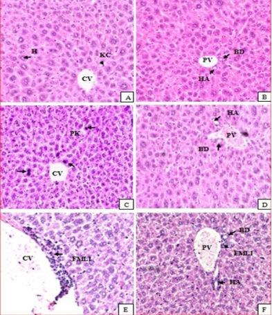
Figure 1. Photomicrographs of sections of liver from control mice (A & B) and, mice treated with 700 mg/kg body weight/day (C & D) and 2100 mg/kg body weight/day (E& F). sections from female mice PK= pyknotic nucleus in hepatocytes in mice treated with 700mg/kg body weight/day (C); FMLI= focal mononuclear lymphocytic cellular infiltration in mice treated with 2100 mg/kg body weight/day (E&F) in central vein and portal area. H= Hepatocytes; BD=Bile duct; HA=Hepatic artery; CV= Central vein; PV=Portal vein; KC=kupperfer cell. (Sections were stained with H&E, X300).
3.3. Effects on Histology of Liver
Histopathological studies of the liver sections in the control group (Figure 1(A) and 1(B)) showed normal appearance of central vein (CV) and hepatic sinusoids (S) lined by endothelial cells (EC) with normal radiating hepatocytes. There was also normal appearance of the portal triad including hepatic portal vein (PV), interlobular bile duct (BD), and branches of hepatic artery (HA). Mice treated with aqueous leaf extracts of M.gracilipes at both doses of 700 mg/kg (Figure 1(C) and 1(D)) and 2100mg/kg (Figure 1(E) and 1(F)) showed normal appearance of the central veins (CV) and hepatic sinusoids(S) lined with endothelial cells (E) with normal radiating hepatocytes. However, some pyknotic nucleuses in hepatocytes were exhibited and perivascular leukocytic cellular infiltration in the central and portal area Figure 1(E) and (F).
3.4. Effects on Histology of Kidneys
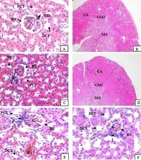
Histopathological studies of the kidneys sections of mice treated with doses of 700 mg/kg (Figure 2(C) and 2(D)) and 2100 mg/kg (Figure 2(E) and 2(F)) showed no significant microscopic/histological changes compared with the controls (Figure 2(A) and 2(B)). In the treated mice of kidney sections revealed normal glomerulus (G), Bowman’s capsule lined with outer parietal layer/squamous cells (SC) and inner visceral layer/podocytes, urinary/bowman’s space (BS), proximal convoluted tubules (PCTs) lined by simple cuboidal epithelium with brush border, distal convoluted tubules (DCTs) lined by simple cuboidal epithelium with more nuclei per cross-section, and macula densa (MD) with taller cells around the vascular pole.
Figure 2. Photomicrographs of sections of kidney of control mice (A & B), mice treated with 700 mg/kg dose/day (C and D) and 2100 mg/kg dose body weight /day (E and F).sections from female mice Mononuclear lymphocytic cellular infiltration in mice treated with 700 mg/kg dose body weight (C), and treated with 2100 mg/kg dose body weight (E&F). DCT=Distal convoluted tubule); PCT=Proximal convoluted tubule; MD=Macula densa; BS=Bowman’s space; G=Glomerulus; BV=Blood Vessles; CA=Cortical area; CMJ=Cortico medullary junction; MA=Medullary area; (Sections were stained with H & E, X300 for A, C, E &F X60 for B&D.
4. Discussion
In toxicological evaluation, biochemical parameters have significant roles as a marker because of their response to clinical signs and symptoms produced by toxicants. Evaluation of hepatic and renal function is major importance to evaluate the toxic properties of extracts and drugs
[18]
Rahman MF, Siddiqui MK, Jamil K. Effects of Vepacide (Azadirachta indica) on asp artate and al anine aminotransferase profiles in a subchronic study with rats. Hum Exp Toxicol. 2001 May 1; 20(5): 243–9.
[18]
. In the present study, all biochemical parameters did not show significant changes.
Measurements of urea and Creatinine levels in the blood are usually performed to evaluate kidney function. Urea is the major nitrogen containing metabolic end product of protein catabolism, and Creatinine is a waste product of muscle energy metabolism. Creatinine and urea concentrations are used for the assessment of renal sufficiency.
Urea is usually increased in acute and chronic renal diseases. Urea clearance falls as the kidney fails and as a result, urea tends to accumulate with failed kidneys that are unable to excrete these substances at normal rate; this will raise blood urea level
[19]
Sonntag O. Tietz Fundamentals of Clinical Chemistry, Fifth Edition. Carl A. Burtis and Edward R. Ashwood, eds. Philadelphia: WB Saunders, 2001, 1091 pp., $75.00. ISBN 0-7216-8634-6. Clin Chem. 2002 Jan 1; 48(1): 213–213.
[20]
Ugwah-Oguejiofor CJ, Okoli CO, Ugwah MO, Umaru ML, Ogbulie CS, Mshelia HE, et al. Acute and sub-acute toxicity of aqueous extract of aerial parts of Caralluma dalzielii N. E. Brown in mice and rats. Heliyon [Internet]. 2019 Jan 29 [cited 2019 Jul 28]; 5(1). Available from:
. In the present study, the mean values of urea have been shown with a slight increment at dose of 700 and 2100mg/kg with none significant with compare to the control. Creatinine is produced and released in to body fluids at a constant rate and its plasma concentration is maintained mainly by glomerular filtration. Consequently, both plasma concentration and its renal clearance have been used as markers of the glomerular filtration rate
[19]
Sonntag O. Tietz Fundamentals of Clinical Chemistry, Fifth Edition. Carl A. Burtis and Edward R. Ashwood, eds. Philadelphia: WB Saunders, 2001, 1091 pp., $75.00. ISBN 0-7216-8634-6. Clin Chem. 2002 Jan 1; 48(1): 213–213.
[19]
. In the current study, the mean amount of Creatinine in treated groups showed slight increment but not significant compared to the control group.
Serum total bilirubin change is caused by a change in the volume of plasma water and a change in the concentration of one or more specific proteins in the plasma. Decrease in the volume of plasma water (hyperproteinemia) is noted in cases of dehydration due to inadequate water intake or excessive water loss, in case of severe vomiting or diarrhea
[19]
Sonntag O. Tietz Fundamentals of Clinical Chemistry, Fifth Edition. Carl A. Burtis and Edward R. Ashwood, eds. Philadelphia: WB Saunders, 2001, 1091 pp., $75.00. ISBN 0-7216-8634-6. Clin Chem. 2002 Jan 1; 48(1): 213–213.
[19]
. In the current study, the amounts of total bilirubin were slightly increased at dose of 700 and 2100 mg/kg compared to control but not statistically significant.
The abnormal elevation of the liver enzymes (ALT and AST) is usually associated with liver damage or change in bile flow. ALT is found primarily in the liver and is the most sensitive marker for liver cell damage. When a cell is damaged, it leaks this enzyme into the blood. AST is found primarily in the red blood cells, cardiac and skeletal muscles, and kidney. AST is not specific to liver as ALT. In this study, the mean values of AST and ALT at dose 700mg/kg and 2100 mg/kg were slightly higher compared with control, but it was not significant. This was supported by the absence of histopathological changes in the liver of treated mice.
The liver and kidneys have fundamental roles in the metabolism and excretion of drugs or plant products. Plant chemicals and their metabolites might result in toxicity or cell damage on target organs
[21]
Debelo N, Afework M. Assessment of Hematological, Biochemical and Histopathological Effects of Acute and Sub-chronic Administration of the Aqueous Leaves Extract of Thymus schimperi in Rats. J Clin Toxicol [Internet]. 2016 [cited 2019 Dec 27]; 06(02). Available from:
Feng Y, Cheung K-F, Wang N, Liu P, Nagamatsu T, Tong Y. Chinese medicines as a resource for liver fibrosis treatment. Chin Med. 2009 Aug 20; 4: 16.
[21, 22]
. In the current histopathological examination of the liver, mice treated with doses of 700 mg/kg and 2100 mg/kg of the aqueous leaves of M. gracilipes showed no change in the microscopic structure of the liver. The general architecture of the liver, appearance of the hepatocytes, the hepatic sinusoids, portal triads, and central veins are normal as compared with controls. The result was also supported by the no adverse effects of the extract in any of the biochemical markers (such as ALT and AST), which showed statistically insignificant changes compared with control group
[23]
Loha M, Mulu A, Abay SM, Ergete W, Geleta B. Acute and Subacute Toxicity of Methanol Extract of Syzygium guineense Leaves on the Histology of the Liver and Kidney and Biochemical Compositions of Blood in Rats [Internet]. Evidence-Based Complementary and Alternative Medicine. 2019 [cited 2019 Dec 27]. Available from:
Feng Y, Cheung K-F, Wang N, Liu P, Nagamatsu T, Tong Y. Chinese medicines as a resource for liver fibrosis treatment. Chin Med. 2009 Aug 20; 4: 16.
[22]
who reported that mice treated at a dose of 200 and 400 mg/kg bodyweight of the aqueous leaves extract of S. guineense showed no histopathological changes compared to control group, whereas the tissue morphology of mice treated with 600mg/kg showed hemorrhagic centrolobular necrosis and fatty cytoplasmic vacuolation of the hepatocytes.
In the histopathological study of the kidney, mice treated with both doses (700 and 2100mg/kg) of the extract showed no significant difference compared to controls. The sections of the kidneys of treated mice showed normal general structure of the kidney and the normal appearance of glomeruli and tubules. The proximal convoluted tubules, distal convoluted tubules, and macula densa are normal. The result was further supported by the values of biochemical parameters of the blood (such as Urea, Creatinine and total Bilirubin/protein), which are main indicators of kidney damage
[23]
Loha M, Mulu A, Abay SM, Ergete W, Geleta B. Acute and Subacute Toxicity of Methanol Extract of Syzygium guineense Leaves on the Histology of the Liver and Kidney and Biochemical Compositions of Blood in Rats [Internet]. Evidence-Based Complementary and Alternative Medicine. 2019 [cited 2019 Dec 27]. Available from:
Feng Y, Cheung K-F, Wang N, Liu P, Nagamatsu T, Tong Y. Chinese medicines as a resource for liver fibrosis treatment. Chin Med. 2009 Aug 20; 4: 16.
[22]
. Who reported absence of difference in tissue morphology between control group and mice treated with the low dose, 200 mg/kg.
5. Conclusion
Sub chronic toxicity study of the aqueous leaves extract of the M.gracilipes did not adversely affect the biochemical parameters of treated mice at both doses and there was no signs of toxicity observed in the histology of kidney and liver sections of treated mice.
Abbreviations
AAU: Addis Ababa University
ALP: Alkaline Phosphatase
ANOVA: Analysis of Variance
ALT: Alanine Transaminase
AST: Aspartate Transaminase
DPX: Dibutyl Phthalate in Xylene
EDTA: Ethylene Diamine Tetra-acetic Acid
EPHI: Ethiopian Public Health Institute
H & E: Haematoxylin and Eosinconcentration
OECD: Organization of Economic Cooperationand Development
SPSS: Statistical Package for Social Science
SEM: standard Error of Mean
WHO: World Health Organization
Acknowledgments
The authors are thankful for the financial support provided by School of Graduate Studies of Addis Ababa University (AAU). And the authors also would like to thank the staff of the Traditional and Modern Medicine Research Directorate at EPHI and Departments of Anatomy, Physiology, pharmacology and Pathology at AAU for their assistance during the study.
Authors’ Contribution
Mengistu Ayele and Minale Fekadie conceived and designed the study; performed the experiments; and analyzed the data. Mekebeb Afework, Eyasu Mekonnen, Asfaw Debella, and Tesfaye Tolessa helped to design the study and supervised the study. Wondwossen Ergete helped to perform histopathological study. Mekebeb Afework and Eyasu Mekonnen drafted and edited the manuscript. All authors approved the final manuscript.
Data Availability
The data used to support the findings of this study are included within the manuscript.
Ethical Approval
Ethics approval for this study was obtained from the Scientific and Ethical Review Committee of the Department of Anatomy, Addis Ababa University.
Conflicts of Interest
The authors declare no conflicts of interest.
References
[1]
Robinson M, Zhang X. THE WORLD MEDICINES SITUATION 2011 TRADITIONAL MEDICINES: GLOBAL SITUATION, ISSUES AND CHALLENGES. In 2011.
[2]
Addis G, Abebe DT, Genebo T, Urga K. Perceptions and practices of modern and traditional health practitioners about traditional medicine in Shirka district, Arsi zone, Ethiopia. In 2002.
[3]
Berhanu A. Book Review: Ethiopian Traditional Medicine: Common Medicinal Plants in Perspective. J Black Stud. 2002 May 1; 32(5): 610–2.
[4]
De Smet PAGM. Traditional pharmacology and medicine in Africa: Ethnopharmacological themes in sub-Saharan art objects and utensils. J Ethnopharmacol. 1998 Nov 1; 63(1): 1–175.
[5]
Gedif T, Hahn H-J. The use of medicinal plants in self-care in rural central Ethiopia. J Ethnopharmacol. 2003 Aug 1; 87(2): 155–61.
[6]
Alves RR, Rosa IM. Biodiversity, traditional medicine and public health: where do they meet? J Ethnobiol Ethnomedicine. 2007 Mar 21; 3(1): 14.
[7]
Azaizeh H, Saad B, Khalil K, Said O. The State of the Art of Traditional Arab Herbal Medicine in the Eastern Region of the Mediterranean: A Review. Evid Based Complement Alternat Med. 2006 Jun; 3(2): 229–35.
[8]
Gedif T, Hahn H-J. Herbalists in Addis Ababa and Butajira, Central Ethiopia: Mode of service delivery and traditional pharmaceutical practice. Ethiop J Health Dev. 2002 Jan 1; 16(2): 183-189–189.
[9]
Chester AH, Yacoub MH. The role of endothelin-1 in pulmonary arterial hypertension. Glob Cardiol Sci Pract. 2014 Jun 18; 2014(2): 62–78.
[10]
Kumaran RS, Jung H, Kim HJ. In vitro screening of taxol, an anticancer drug produced by the fungus, Colletotrichum capsici. Eng Life Sci. 2011; 11(3): 264–71.
[11]
Schümann J, Prockl J, Kiemer AK, Vollmar AM, Bang R, Tiegs G. Silibinin protects mice from T cell-dependent liver injury. J Hepatol. 2003 Sep 1; 39(3): 333–40.
[12]
Angerhofer CK. Herbal Medicines, a Guide for Healthcare Professionals, Second Edition By Joanne Barnes (University of London), Linda A. Anderson (Medicines Control Agency, London), and J. David Phillipson (University of London). Pharmaceutical Press, London, UK. 2002. xiv + 530 pp. 18.5 × 24.5 cm. $59.95. ISBN 0-85369-474-5. J Nat Prod. 2002 Dec 1; 65(12): 1964–1964.
[13]
Mahomoodally MF. Traditional Medicines in Africa: An Appraisal of Ten Potent African Medicinal Plants [Internet]. Evidence-Based Complementary and Alternative Medicine. 2013 [cited 2019 Dec 27]. Available from:
Adebayo JO, Yakubu MT, Egwim EC, Owoyele VB, Enaibe BU. Effect of ethanolic extract of Khaya senegalensis on some biochemical parameters of rat kidney. J Ethnopharmacol. 2003 Sep 1; 88(1): 69–72.
[15]
Geyid A, Abebe D, Debella A, Makonnen Z, Aberra F, Teka F, et al. Screening of some medicinal plants of Ethiopia for their anti-microbial properties and chemical profiles. J Ethnopharmacol. 2005 Jan 1.
[16]
Test No. 425: Acute Oral Toxicity: Up-and-Down Procedure - en - OECD [Internet]. [cited 2019 Jul 14]. Available from:
Saleem U, Amin S, Ahmad B, Azeem H, Anwar F, Mary S. Acute oral toxicity evaluation of aqueous ethanolic extract of Saccharum munja Roxb. roots in albino mice as per OECD 425 TG. Toxicol Rep. 2017 Oct 31; 4: 580–5.
[18]
Rahman MF, Siddiqui MK, Jamil K. Effects of Vepacide (Azadirachta indica) on asp artate and al anine aminotransferase profiles in a subchronic study with rats. Hum Exp Toxicol. 2001 May 1; 20(5): 243–9.
[19]
Sonntag O. Tietz Fundamentals of Clinical Chemistry, Fifth Edition. Carl A. Burtis and Edward R. Ashwood, eds. Philadelphia: WB Saunders, 2001, 1091 pp., $75.00. ISBN 0-7216-8634-6. Clin Chem. 2002 Jan 1; 48(1): 213–213.
[20]
Ugwah-Oguejiofor CJ, Okoli CO, Ugwah MO, Umaru ML, Ogbulie CS, Mshelia HE, et al. Acute and sub-acute toxicity of aqueous extract of aerial parts of Caralluma dalzielii N. E. Brown in mice and rats. Heliyon [Internet]. 2019 Jan 29 [cited 2019 Jul 28]; 5(1). Available from:
Debelo N, Afework M. Assessment of Hematological, Biochemical and Histopathological Effects of Acute and Sub-chronic Administration of the Aqueous Leaves Extract of Thymus schimperi in Rats. J Clin Toxicol [Internet]. 2016 [cited 2019 Dec 27]; 06(02). Available from:
Feng Y, Cheung K-F, Wang N, Liu P, Nagamatsu T, Tong Y. Chinese medicines as a resource for liver fibrosis treatment. Chin Med. 2009 Aug 20; 4: 16.
[23]
Loha M, Mulu A, Abay SM, Ergete W, Geleta B. Acute and Subacute Toxicity of Methanol Extract of Syzygium guineense Leaves on the Histology of the Liver and Kidney and Biochemical Compositions of Blood in Rats [Internet]. Evidence-Based Complementary and Alternative Medicine. 2019 [cited 2019 Dec 27]. Available from:
Ayele, M., Afework, M., Makonnen, E., Ergete, W., Debella, A., et al. (2024). Sub Chronic Toxicity Study of Aqueous Leaves Extract of Maytenus Gracilipes on Some Biochemical Parameters and Histopathology of Liver and Kidneys in Mice. American Journal of Bioscience and Bioengineering, 12(2), 24-31. https://doi.org/10.11648/j.bio.20241202.11
Ayele, M.; Afework, M.; Makonnen, E.; Ergete, W.; Debella, A., et al. Sub Chronic Toxicity Study of Aqueous Leaves Extract of Maytenus Gracilipes on Some Biochemical Parameters and Histopathology of Liver and Kidneys in Mice. Am. J. BioSci. Bioeng.2024, 12(2), 24-31. doi: 10.11648/j.bio.20241202.11
Ayele M, Afework M, Makonnen E, Ergete W, Debella A, et al. Sub Chronic Toxicity Study of Aqueous Leaves Extract of Maytenus Gracilipes on Some Biochemical Parameters and Histopathology of Liver and Kidneys in Mice. Am J BioSci Bioeng. 2024;12(2):24-31. doi: 10.11648/j.bio.20241202.11
@article{10.11648/j.bio.20241202.11,
  author = {Mengistu Ayele and Mekebeb Afework and Eyasu Makonnen and Wondwosen Ergete and Asfaw Debella and Tesfaye Tolessa and Minale Fekadie},
  title = {Sub Chronic Toxicity Study of Aqueous Leaves Extract of Maytenus Gracilipes on Some Biochemical Parameters and Histopathology of Liver and Kidneys in Mice
},
  journal = {American Journal of Bioscience and Bioengineering},
  volume = {12},
  number = {2},
  pages = {24-31},
  doi = {10.11648/j.bio.20241202.11},
  url = {https://doi.org/10.11648/j.bio.20241202.11},
  eprint = {https://article.sciencepublishinggroup.com/pdf/10.11648.j.bio.20241202.11},
  abstract = {Introduction: Traditional medicine is an ancient medical practice that is still widely used in prevention and treatment of various health problems worldwide, including Ethiopia. M.gracilipes is one of medicinal plant in Ethiopia used for treatment of various ailments still is very common. This study evaluated the sub-chronic toxic effects of M. gracilipes aqueous leaves extract on biochemical parameters and histopathology of liver and kidneys. Methods: For sub-chronic toxicity study a total of 30 mice were used, three groups (I–III) of mice (10 animals each) were used. Group I served as control and received a vehicle while groups II and III daily administered with 700 and 2100 mg/kg extract respectively orally by using oral gavage for 90 days. At the end of the experiment, the mice were sacrificed by using diethyl ether; blood was collected for assessing biochemical parameters and histopathological evaluations on liver and kidneys were performed. Results: Sub chronic treatment of extract for 90 days, at 700 and 2100 mg/kg body weight did not induce any sign of illness and /or death and had no adverse effect on biochemical parameters and blood parameters. Liver and kidney sections also revealed normal architecture, except some pyknotic nuclei and focal mononuclear leukocytic infiltrations observed in some of the liver and kidney tissues at higher dose (2100mg/kg). Conclusion: The results of this sub chronic toxicity study showed that M. gracilipes aqueous leaves extract is safe at daily doses of 700 mg/kg body weight, even when taken for longer period. At higher doses, however, the extract may induce mild hepatorenal toxicity.
},
 year = {2024}
}
TY  - JOUR
T1  - Sub Chronic Toxicity Study of Aqueous Leaves Extract of Maytenus Gracilipes on Some Biochemical Parameters and Histopathology of Liver and Kidneys in Mice

AU  - Mengistu Ayele
AU  - Mekebeb Afework
AU  - Eyasu Makonnen
AU  - Wondwosen Ergete
AU  - Asfaw Debella
AU  - Tesfaye Tolessa
AU  - Minale Fekadie
Y1  - 2024/04/17
PY  - 2024
N1  - https://doi.org/10.11648/j.bio.20241202.11
DO  - 10.11648/j.bio.20241202.11
T2  - American Journal of Bioscience and Bioengineering
JF  - American Journal of Bioscience and Bioengineering
JO  - American Journal of Bioscience and Bioengineering
SP  - 24
EP  - 31
PB  - Science Publishing Group
SN  - 2328-5893
UR  - https://doi.org/10.11648/j.bio.20241202.11
AB  - Introduction: Traditional medicine is an ancient medical practice that is still widely used in prevention and treatment of various health problems worldwide, including Ethiopia. M.gracilipes is one of medicinal plant in Ethiopia used for treatment of various ailments still is very common. This study evaluated the sub-chronic toxic effects of M. gracilipes aqueous leaves extract on biochemical parameters and histopathology of liver and kidneys. Methods: For sub-chronic toxicity study a total of 30 mice were used, three groups (I–III) of mice (10 animals each) were used. Group I served as control and received a vehicle while groups II and III daily administered with 700 and 2100 mg/kg extract respectively orally by using oral gavage for 90 days. At the end of the experiment, the mice were sacrificed by using diethyl ether; blood was collected for assessing biochemical parameters and histopathological evaluations on liver and kidneys were performed. Results: Sub chronic treatment of extract for 90 days, at 700 and 2100 mg/kg body weight did not induce any sign of illness and /or death and had no adverse effect on biochemical parameters and blood parameters. Liver and kidney sections also revealed normal architecture, except some pyknotic nuclei and focal mononuclear leukocytic infiltrations observed in some of the liver and kidney tissues at higher dose (2100mg/kg). Conclusion: The results of this sub chronic toxicity study showed that M. gracilipes aqueous leaves extract is safe at daily doses of 700 mg/kg body weight, even when taken for longer period. At higher doses, however, the extract may induce mild hepatorenal toxicity.

VL  - 12
IS  - 2
ER  -